Report on the WRHI Lecture 2 (12th July, 2017)
Events
Report on the WRHI Lecture 2 (12th July, 2017)
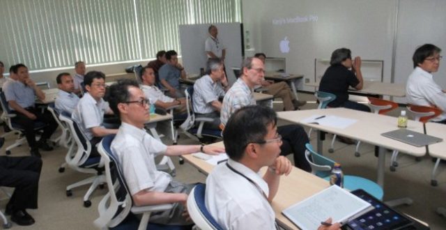
On the afternoon of 12th July, the second WRHI Lecture session was held in the Open Communication Space of the R2 Building. This session saw an increase in participant numbers, reflecting the popularity of these lecture and establishment of the program. This time, 3 researchers from abroad, invited to Tokyo Tech through the WRHI program, gave lectures on their own research, hosted by Professor Masaki Higashi of the Laboratory for Materials and Structures.
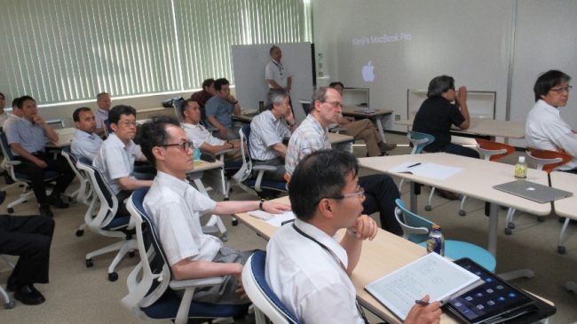 Attending academics (left)
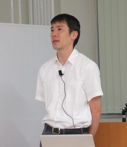
 and Professor Azuma (right), who chaired the session
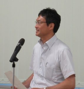
The first lecture was delivered by Dr. Alexander I May, who works in the Ohsumi laboratory and was invited from Monash University in Australia. He gave a lecture entitled “The physiological role of autophagy in metabolically transitioning cells”.
He began by explaining the life cycle, environmental challenges and physiology of budding yeast cells, which are used as a model organism in his work (Saccharomyces cerevisiae). Yeast have been known since ancient times to ferment alcohol by metabolizing sugar. Dr. May then discussed the role autophagy plays as a response to environmental challenges. Autophagy is a biological recycling system that allows cells to *eat* (degrade) their own constituents in the absence of external nutrients. Dr. May compares normal cells with cells defective for autophagy, and has found that cells lacking autophagy are characterized by a defect in growth. However, it was confirmed that supplementation of certain kinds of amino acids can relieve the growth defect of this mutant. The significance of amino acids in this circumstance was linked to the system that synthesizes proteins in mitochondria, which are in important center in cell metabolism. This lecture provided an insight into the latest research in autophagy, and includes new and complicated processes and methodologies. This work promises to provide insights into the relatively unexplored physiological role of autophagy.
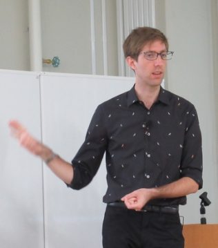
Next, Dr. Kenji Suzuki gave a lecture entitled “Artificial Intelligence in Medical Imaging”. Dr. Suzuki studied computational intelligence (or artificial intelligence) in medical image analysis at University of Chicago for 13 years and then at Illinois Institute of Technology for 3 years.
First, Dr. Suzuki explained what computer-aided diagnosis is by analogy to the popular children’s book, “Where’s Waldo”. In computer-aided diagnosis, artificial intelligence (AI) detects lesion candidates in medical images; and then, a radiologist makes final diagnosis by taking into account computer suggestions as a “second opinion”. Machine learning plays a central role in AI. There are a large variety of shapes and patterns of lesions and organs in medical images, which require complex models to represent. A complex model has a large number of parameters to adjust. It is generally difficult to determine such a large number of parameters by hands. Thus, machine learning that automatically and reasonably determines them by “learning from examples (or data)” is essential in AI in computer-aided diagnosis. In conventional approaches, AI first segments lesions from medical images; and then, features are extracted from the segmented lesions. The extracted features are entered into a machine-learning model to classify lesions into known classes. However, conventional machine-learning approaches did not reach at the diagnostic performance of radiologists. In 1996, Dr. Suzuki and his colleagues invented and developed novel machine learning that learns images directly. Dr. Suzuki and his colleagues at University of Chicago advanced his machine learning and applied it to computer-aided diagnosis in 2003. This new, unique machine-learning framework learns images directly, like children learn objects directly by seeing objects as images. With this technology, the performance of AI was improved substantially. This class of new AI is called “child AI”, whereas the class of the conventional AI approaches that use features is called “adult AI”, because adult people explain an object by features or words. Very recently starting in around 2013, a similar class of method called deep learning obtained enthusiastic attention and became the hottest topic in many fields in industry and academia. “Child AI” with deep learning is expected to improve the performance of computer-aided diagnostic systems dramatically and to revolutionize medicine. The new “child AI” framework that Dr. Suzuki pioneered to develop is highly versatile and thus applicable to many fields including medicine.
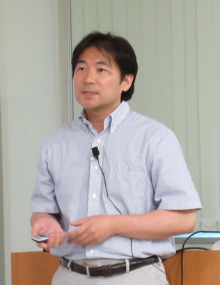
The last lecture was “Intracellular organelle dynamics during cell division cycle” by Dr. Yui Jin invited to the Ohsumi laboratory from Life Sciences Institute, University of Michigan.
The organelle present inside the cell consists of several structures such as nucleus, mitochondria, vacuole, Golgi apparatus, endosome and so on. The topic is to explain what kind of phenomena are occurring in cell organelles during cell division. When cell division begins, daughter cells protruding from the mother cell gradually become larger. And after cell division the daughter cells will have the same organelle as the mother cell. It is a protein called myosin that makes this possible, and it plays a role in carrying various structures of cellular organelles in dividing cells. Although cell division progressed in this way, it was also found that when cell fails to transport of the vacuoles to the daughter cell, the vacuoles are newly synthesized in the daughter cell, and then the cell division will be occurred. This research is a steady basic research in the field of life sciences that will unravel phenomena one by one, but I would like to focus on how to contribute for society in the future.
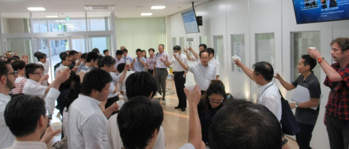
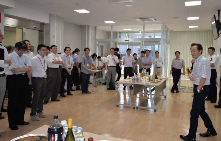
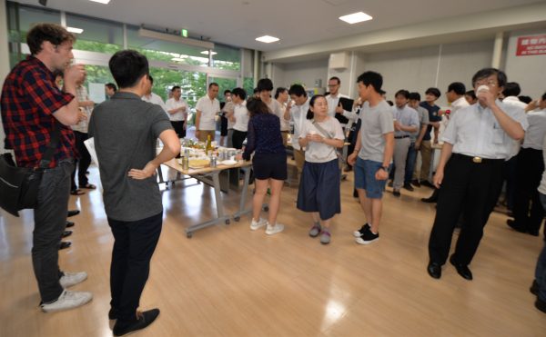
After some lively discussion, the session was concluded successfully. The meeting then adjourned to a party attended by faculty members and students from the Institute of Innovative Research in the adjacent lobby began. Released from the focused atmosphere of the lecture session, the participants enjoyed an early summer’s evening fully with plenty of beer.